Brain Cancer and Brain Tumours

Understanding the brain

The main parts of the brain include:

The cerebrum. This is divided into the right side (right hemisphere) which controls the left side of the body, and the left hemisphere which controls the right side of the body. Each hemisphere is divided into various subsections, the main divisions being the frontal lobe, temporal lobe, parietal lobe and occipital lobe. The cerebrum is also where you think and store your memory.

The cerebellum. This lies behind and below the cerebrum. One of its main functions is to help control balance and co-ordination.

The brain stem. This helps to control basic bodily functions such as the heartbeat, breathing and blood pressure. Nerves from the cerebrum also pass through the brainstem to the spinal cord.

The meninges. These are thin layers of tissue which separate the skull from the brain. The outer layer next to the skull is called the dura. The next layer is called the arachnoid. Under the arachnoid tissue is the cerebrospinal fluid (CSF) which bathes the brain and spinal cord.

The pituitary gland. This releases various hormones into the bloodstream.

The main type of cell in the brain is called a neuron. There are millions of neurons in the brain. Neurons have long thin nerve fibres which enable them to send messages to other parts of the brain and down the spinal cord to all parts of the body. The brain also contains glial cells. These provide support, nourishment and protection for neurons. There are various types of glial cells, including astrocytes, oligodendrocytes and ependymal cells.

The original site where a tumour first develops is called a primary tumour. Cancerous (malignant) tumours may also spread to other parts of the body to form secondary tumours (metastases). These secondary tumours may then grow, invade and damage nearby tissues and spread again.

Primary malignant brain tumours

A primary malignant brain tumour is a cancer which arises from a cell within the brain. The cells of the tumour grow into and damage normal brain tissue. Also, like non-cancerous (benign) brain tumours, they can increase the pressure inside the skull. However, unlike most other types of malignant tumours, primary brain tumours rarely spread (metastasise) to other parts of the body.

There are various types of primary malignant brain tumour. The different types develop from different types of cell in the brain. As a general guide, each type is graded on a scale of 1-4. Grade 1 and grade 2 tumours are said to be low-grade; grade 3 and grade 4 are termed high-grade. The higher the grade, the more aggressive the tumour tends to be and the faster it tends to grow. The treatment options and outlook (prognosis) can vary depending on the type and grade of the tumour.

Secondary malignant brain tumours

A secondary malignant brain tumour means that a cancer which started in another part of the body has spread to the brain. Many types of cancer can spread to the brain. The most common types that do this are cancers of the breastlungbowelkidney and skin (melanoma).

See the separate leaflet called Cancer for more general information about cancer

There are many types of non-cancerous (benign) brain tumours and primary cancerous (malignant) brain tumours. Many are very rare. The following is a brief description of the most common types.

Meningioma

Meningiomas are usually benign. They grow from cells in the tissues that surround the brain (the meninges).

Medulloblastoma

These are high-grade malignant tumours that grow in the cerebellum. They are uncommon in adults but are one of the two most common brain tumours in children. The other is astrocytoma in the cerebellum.

Gliomas

These are malignant primary brain tumours that arise from glial cells. There are various types, depending on the cell of origin – for example:

  • Astrocytomas (originating from astrocyte cells.) There are various types of astrocytoma. They include:
    • Low-grade astrocytomas.
    • Anaplastic astrocytoma. This is a high-grade tumour.
    • Glioblastoma multiforme. This is a high-grade tumour which tends to grow quite quickly. It is the most common type of primary malignant brain tumour in adults.
  • Oligodendrogliomas (originating from oligodendrocytes). These can vary in their grade.
  • Ependymoma (originating from ependymal cells). These are rare but are usually low-grade.

Primitive neuroectodermal tumours (PNETs)

These are very similar to medulloblastomas and mainly occur in children.

Pituitary tumours

There are various types of tumour which arise from the different cells in the pituitary gland. They tend to be benign. However, the cells of the tumour may produce large quantities of hormones which can cause various symptoms. As they grow, they may also cause pressure symptoms. The nerves of sight (optic nerves) are near to the pituitary gland and so a growing pituitary gland tumour may press on an optic nerve and affect vision.

Acoustic neuroma

An acoustic neuroma (schwannoma) is a benign tumour which arises from Schwann cells which cover the nerve that goes to the ear. Symptoms can include deafness on the affected side and dizziness with a spinning sensation (vertigo).

Other

There are various other rare types of benign and primary malignant brain tumours.

The cause of most non-cancerous (benign) brain tumours and primary cancerous (malignant) brain tumours is not known.

Genetic factors may be a risk for some people – perhaps in about 1 in 20 cases. For example, people with the hereditary diseases called neurofibromatosis type 1, Turcot’s syndrome, Li-Fraumeni cancer syndrome, and tuberous sclerosis have a higher-than-average risk of developing a glioma. When people with these diseases develop a glioma, it tends to occur in childhood or early adult life. However, these cases are only a small proportion of all glioma tumours.

Note:

  • Most gliomas occur in older adults and inherited (genetic) factors are not thought to be involved.
  • Previous radiotherapy to the brain is thought to increase the risk of a brain tumour.
  • There is no strong evidence that using mobile phones increases the risk of brain tumours.
  • Secondary (metastatic) brain tumours arise from various cancers of the body. These have various causes. See the separate leaflets about these other cancers.

Non-cancerous (benign) brain tumours and cancerous (malignant) primary brain tumours are uncommon. Overall they occur in about 12 in 100,000 people each year.

The most common types in adults are benign meningioma and a glioma called glioblastoma multiforme. Some types are very rare.

Brain tumours can occur at any age. Some types (such as medulloblastoma) are more common in children and some are more common in adults. Generally, the tumours that tend to occur in adults become more common with increasing age.

Secondary (metastatic) brain tumours are more common than benign brain tumours and malignant primary brain tumours.

General symptoms

Early symptoms may include headaches and feeling sick. These are due to increased pressure within the skull (raised intracranial pressure). These symptoms may come and go at first and tend to be worse in the morning. Coughing, sneezing and stooping may make the headaches worse. Epileptic seizures (convulsions) sometimes occur. Increasing drowsiness may occur as the tumour enlarges.

Note: most people who have headaches or epilepsy do not have a brain tumour.

Symptoms due to the location in the brain

As a tumour grows it can damage the nearby brain tissue. The functions of the different parts of the body are controlled by different parts of the brain. Therefore, the symptoms vary from case to case, depending on which part of the brain is affected and on the size of the affected area. For example, one or more of the following may develop:

These symptoms tend to develop gradually.

A doctor will examine you if a brain tumour is suspected from the symptoms. This will include checking on the functions of the brain and nerves (movements, reflexes, vision, etc).

A magnetic resonance imaging (MRI) scan or computerised tomography (CT) scan of the head is the common test done to confirm or rule out the presence of a brain tumour. See the separate leaflets called MRI Scan and CT Scan for more details. If a tumour is identified, further more detailed scans and tests may be done. For example, a PET scan or a cerebral angiogram are sometimes done to obtain more information about the tumour.

small tissue sample (a biopsy) may be needed to be sure of the type of tumour. The sample is then examined under the microscope to look for abnormal cells. To obtain a biopsy from a brain tumour you need to have a small operation, usually done under anaesthetic. A small hole is bored in the skull to allow a fine needle through to obtain a small sample of tissue. By examining the cells obtained by the biopsy, the exact type of tumour can be identified. If it is cancerous (malignant), the tumour grade can be determined (see above).

Blood tests and other tests on other parts of the body may be done if the tumour is thought to be a secondary (metastatic) tumour. For example, it is quite common for a lung cancer to spread to the brain. Therefore, a chest X-ray may be done if this is suspected. Various hormone tests may be done if a pituitary gland tumour is suspected. 

The main treatments used for brain tumours are surgery, chemotherapy, radiotherapy and medication to control symptoms such as seizures. The treatment or combination of treatments advised in each case depends on various factors – for example:

  • The type of brain tumour.
  • The grade of the tumour if it is cancerous (malignant).
  • The exact site of the tumour.
  • Your general health.

Surgery

Surgery is often the main treatment for non-cancerous (benign) brain tumours and primary cancerous (malignant) tumours. The aim of surgery is to remove the tumour (or even some of the tumour) whilst doing as little damage as possible to the normal brain tissue. Your specialist will advise on whether surgery is a possible option.

Radiotherapy

Radiotherapy is a treatment which uses high-energy beams of radiation which are focused on cancerous tissue. This kills cancer cells or stops cancer cells from multiplying. See the separate leaflet called Radiotherapy for more details.

Radiotherapy is sometimes used instead of surgery when an operation is not possible for a malignant brain tumour. Sometimes it is used in addition to surgery if it is not possible to remove all the tumour with surgery or to kill cancerous cells which may be left behind following surgery.

Chemotherapy

Chemotherapy is a treatment which uses anti-cancer medicines to kill cancer cells, or to stop them from multiplying. See the separate leaflet called Chemotherapy for more details. It may be used in addition to other treatments such as surgery or radiotherapy; again, depending on various factors such as the type of tumour.

Medication to control symptoms

If you have seizures caused by the tumour then anticonvulsant medication will usually control the seizures. Painkillers may be needed to ease any headaches. Steroid medication is also commonly used to reduce inflammation around a brain tumour. This reduces the pressure inside the skull, which helps to ease headaches and other pressure symptoms.

You should have a full discussion with a specialist who knows your case. They will be able to give the pros and cons, likely success rate, possible side-effects and other details about the possible treatment options for your type of brain tumour.

You should also discuss with your specialist the aims of treatment. For example:

  • In some cases, treatment aims for a cure. If a benign tumour can be removed by surgery then a cure is likely. The chance of a cure for malignant tumours varies, depending on the type of tumour, grade and other factors such as the location in the brain. Note: when dealing with malignant tumours, doctors tend to use the word remission rather than the word cured. Remission means there is no evidence of cancer following treatment. If you are in remission, you may be cured. However, in some cases a cancer returns months or years later. This is why doctors are sometimes reluctant to use the word cured.
  • In some cases, treatment aims to control the cancer. If a cure is not realistic, with treatment it may be possible to limit the growth or spread of the cancer so that it progresses less rapidly. This may keep you free of symptoms for some time.
  • In some cases, treatment aims to ease symptoms (palliative treatment). For example, if a cancer is advanced then you may require painkillers or other treatments to help keep you free of pain or other symptoms. Some treatments may be used to reduce the size of a cancer, which may ease symptoms such as pain.

It is difficult to give an overall outlook (prognosis). Every case is different. For example, if you have a non-cancerous (benign) meningioma which is in a suitable place for surgery, the outlook is excellent.

For primary cancerous (malignant) brain tumours, the outlook will vary, depending on the type, grade and location in the brain.

The outlook is often poor if you have a secondary (metastatic) malignant brain tumour.

The treatment of cancer is a developing area of medicine. New treatments continue to be developed and the information on outlook above is very general. The specialist who knows your case can give more accurate information about your particular outlook and how well your type and stage of cancer is likely to respond to treatment. 

Chiari malformation

Overview

Chiari malformation

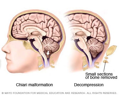
Chiari malformation (kee-AH-ree mal-for-MAY-shun) is a condition in which brain tissue extends into your spinal canal. It occurs when part of your skull is abnormally small or misshapen, pressing on your brain and forcing it downward.

Chiari malformation is uncommon, but increased use of imaging tests has led to more frequent diagnoses.

Doctors categorize Chiari malformation into three types, depending on the anatomy of the brain tissue that is displaced into the spinal canal and whether developmental abnormalities of the brain or spine are present.

Chiari malformation type I develops as the skull and brain are growing. As a result, signs and symptoms may not occur until late childhood or adulthood. The pediatric forms, Chiari malformation type II and type III, are present at birth (congenital).

Treatment of Chiari malformation depends on the form, severity and associated symptoms. Regular monitoring, medications and surgery are treatment options. In some cases, no treatment is needed.

Symptoms

Many people with Chiari malformation have no signs or symptoms and don’t need treatment. Their condition is detected only when tests are performed for unrelated disorders. However, depending on the type and severity, Chiari malformation can cause a number of problems.

The more common types of Chiari malformation are:

  • Type I
  • Type II

Although these types are less serious than the more rare pediatric form, type III, signs and symptoms still can be life disrupting.

Chiari malformation type I

In Chiari malformation type I, signs and symptoms usually appear during late childhood or adulthood.

Headaches, often severe, are the classic symptom of Chiari malformation. They generally occur after sudden coughing, sneezing or straining. People with Chiari malformation type I can also experience:

  • Neck pain
  • Unsteady gait (problems with balance)
  • Poor hand coordination (fine motor skills)
  • Numbness and tingling of the hands and feet
  • Dizziness
  • Difficulty swallowing, sometimes accompanied by gagging, choking and vomiting
  • Speech problems, such as hoarseness

Less often, people with Chiari malformation may experience:

  • Ringing or buzzing in the ears (tinnitus)
  • Weakness
  • Slow heart rhythm
  • Curvature of the spine (scoliosis) related to spinal cord impairment
  • Abnormal breathing, such as central sleep apnea, which is when a person stops breathing during sleep

Chiari malformation type II

In Chiari malformation type II, a greater amount of tissue extends into the spinal canal compared with Chiari malformation type I.

The signs and symptoms can include those related to a form of spina bifida called myelomeningocele that nearly always accompanies Chiari malformation type II. In myelomeningocele, the backbone and the spinal canal haven’t closed properly before birth.

Signs and symptoms may include:

  • Changes in breathing pattern
  • Swallowing problems, such as gagging
  • Quick downward eye movements
  • Weakness in arms

Chiari malformation type II is usually noted with ultrasound during pregnancy. It may also be diagnosed after birth or in early infancy.

Chiari malformation type III

In one of the most severe types of the condition, Chiari malformation type III, a portion of the lower back part of the brain (cerebellum) or the brainstem extends through an abnormal opening in the back of the skull. This form of Chiari malformation is diagnosed at birth or with an ultrasound during pregnancy.

This type of Chiari malformation has a higher mortality rate and may also cause neurological problems.

When to see a doctor

If you or your child has any of the signs and symptoms that may be associated with Chiari malformation, see your doctor for an evaluation.

Because many symptoms of Chiari malformation can also be associated with other disorders, a thorough medical evaluation is important.

Causes

Chiari malformation type I occurs when the section of your skull containing a part of your brain (cerebellum) is too small or is deformed, thus putting pressure on and crowding your brain. The lower part of the cerebellum (tonsils) is displaced into your upper spinal canal.

Chiari malformation type II is nearly always associated with a form of spina bifida called myelomeningocele.

When the cerebellum is pushed into your upper spinal canal, it can interfere with the normal flow of cerebrospinal fluid that protects your brain and spinal cord.

This impaired circulation of cerebrospinal fluid can lead to the blockage of signals transmitted from your brain to your body, or to a buildup of spinal fluid in the brain or spinal cord.

Alternatively, the pressure from the cerebellum on the spinal cord or lower brainstem can cause neurological signs or symptoms.

Risk factors

There’s some evidence that Chiari malformation runs in some families. However, research into a possible hereditary component is still in its early phase.

Complications

In some people, Chiari malformation can become a progressive disorder and lead to serious complications. In others, there may be no associated symptoms, and no intervention is necessary. The complications associated with this condition include:

  • Hydrocephalus. An accumulation of excess fluid within your brain (hydrocephalus) may require placement of a flexible tube (shunt) to divert and drain the cerebrospinal fluid to another area of your body.
  • Spina bifida. Spina bifida, a condition in which your spinal cord or its covering isn’t fully developed, may occur in Chiari malformation. Part of the spinal cord is exposed, which can cause serious conditions such as paralysis. People with Chiari malformation type II usually have a form of spina bifida called myelomeningocele.
  • Syringomyelia. Some people with Chiari malformation also develop a condition called syringomyelia, in which a cavity or cyst (syrinx) forms within the spinal column.
  • Tethered cord syndrome. In this condition, your spinal cord attaches to your spine and causes your spinal cord to stretch. This can cause serious nerve and muscle damage in your lower body.

Diagnosis

To diagnose your condition, your doctor will review your medical history and symptoms and conduct a physical examination.

Your doctor will also order imaging tests to diagnose your condition and determine its cause. Tests may include:

  • Magnetic resonance imaging (MRI). An MRI is often used to diagnose Chiari malformation. An MRI uses powerful radio waves and magnets to create a detailed view of your body.This safe, painless test produces detailed 3D images of structural abnormalities in your brain that may be contributing to your symptoms. It can also provide images of your cerebellum and determine whether it extends into your spinal canal.An MRI can be repeated over time, and it can be used to monitor the progression of your disorder.
  • Computerized tomography (CT) scan. Your doctor may recommend other imaging techniques such as a CT scan.A CT scan uses X-rays to obtain cross-sectional images of your body. A CT scan can help to reveal brain tumors, brain damage, bone and blood vessel abnormalities, and other conditions.

Treatment

Treatment for Chiari malformation depends on the severity and the characteristics of your condition.

If you have no symptoms, your doctor likely will recommend no treatment other than monitoring with regular examinations and MRIs.

When headaches or other types of pain are the primary symptom, your doctor may recommend pain medication.

Reducing pressure with surgery

Doctors usually treat symptomatic Chiari malformation with surgery. The goal is to stop the progression of changes in the anatomy of your brain and spinal canal, as well as ease or stabilize your symptoms.

When successful, surgery can reduce pressure on your cerebellum and spinal cord, and restore the normal flow of spinal fluid.

In the most common surgery for Chiari malformation, called posterior fossa decompression, your surgeon removes a small section of bone in the back of your skull, relieving pressure by giving your brain more room.

In many cases, the covering of your brain, called the dura mater, may be opened. Also, a patch may be sewn in place to enlarge the covering and provide more room for your brain. This patch may be an artificial material, or it could be tissue harvested from another part of your body.

Your doctor may also remove a small portion of the spinal column to relieve pressure on your spinal cord and allow more space for the spinal cord.

The surgical technique may vary, depending on whether a fluid-filled cavity (syrinx) is present, or if you have fluid in your brain (hydrocephalus). If you have a syrinx or hydrocephalus, you may need a tube (shunt) to drain the excess fluid.

Surgical risks and follow-up

Surgery involves risks, including the possibility of infection, fluid in your brain, cerebrospinal fluid leakage or problems with wound healing. Discuss the pros and cons with your doctor when deciding whether surgery is the most appropriate treatment for you.

The surgery reduces symptoms in most people, but if nerve injury in the spinal canal has already occurred, this procedure won’t reverse the damage.

After the surgery, you’ll need regular follow-up examinations with your doctor, including periodic imaging tests to assess the outcome of surgery and the flow of cerebrospinal fluid.

Preparing for your appointment

You’re likely to start by seeing your family doctor. However, when you call to set up an appointment, you may be referred to a doctor trained in brain and nervous system conditions (neurologist).

Because appointments can be brief, and because there’s often a lot to talk about, it’s a good idea to be well prepared for your appointment. Here’s some information to help you get ready for your appointment, and know what to expect from your doctor.

What you can do

  • Be aware of any pre-appointment restrictions. At the time you make the appointment, be sure to ask if there’s anything you need to do in advance.
  • Write down any symptoms you’re experiencing, including any that may seem unrelated to the reason for which you scheduled the appointment. For example, even though your primary complaint may be headaches, your doctor will want to know about any changes you may have noticed in your vision, speech or coordination.
  • Write down key personal information, including any major stresses and recent life changes.
  • Make a list of your key medical information, including other conditions you’re being treated for and the names of the medications that you’re taking.
  • Take a family member or friend along, if possible. Sometimes it can be difficult to recall all the information provided to you during an appointment. Someone who accompanies you may remember something that you missed or forgot.
  • Write down questions to ask your doctor.

Prepare a list of questions so that you can make the most of your limited time with your doctor. List your questions from most important to least important in case time runs out. For Chiari malformation, some basic questions to ask your doctor include:

  • What is likely causing my symptoms or condition?
  • Other than the most likely cause, what are possible causes for my symptoms or condition?
  • What kinds of tests do I need?
  • Do I need treatment?
  • If you don’t think I need to be treated now, how will you monitor me for changes in my condition?
  • If you recommend surgery, what should I expect from my recovery?
  • What is the risk of complications from surgery?
  • What is my long-term prognosis after surgery?
  • I have other health conditions. How can I best manage them together?
  • Are there any restrictions that I need to follow?
  • Should I see a specialist? What will that cost, and will my insurance cover seeing a specialist?
  • Are there any brochures or other printed material that I can take home with me? What websites do you recommend visiting?

In addition to the questions that you’ve prepared to ask your doctor, don’t hesitate to ask questions during your appointment at any time that you don’t understand something.

What to expect from your doctor

Your doctor is likely to ask you a number of questions. Being ready to answer them may reserve time to go over any points you want to spend more time on. Your doctor may ask:

  • When did you first begin experiencing symptoms?
  • Have your symptoms been continuous or occasional?
  • If you experience head and neck pain, is it made worse by sneezing, coughing or straining?
  • How severe is your head and neck pain?
  • Have you noticed any change in your coordination, including problems with balance or with hand coordination?
  • Do your hands and feet feel numb or do they tingle?
  • Have you developed any difficulty swallowing?
  • Do you experience episodes of dizziness or faintness? Have you ever passed out?
  • Have you developed any problems with your eyes and ears, such as blurred vision or a ringing or buzzing in your ears?
  • Have you had problems with bladder control?
  • Has anyone ever noticed that you stop breathing during sleep?
  • Have you been taking pain relievers or using other approaches to relieve your discomfort? Does anything seem to work?
  • Do you have any additional symptoms, such as hearing loss, fatigue, or changes in your bowel habits or appetite?
  • Have you been diagnosed with any other health conditions?
  • Has anyone in your family been diagnosed with Chiari malformation?

Chiari malformation

My 29yr old daughter suffers from the above neurological condition and has done for sometime now.

I would like to write this blog as a way of support for her because as I type this she is currently in a hospital in Edinburgh talking with a neurosurgeon she will probably get some tests like an MRI done.

My daughter trained in hairdressing & beauty and unfortunately Chiari malformation is preventing her from doing her job.

So I hope we get some positive results from todays visit!

For information on Chiari malformation click the link below

https://www.nhs.uk/conditions/chiari-malformation/

Epilepsy in Pets

What is Epilepsy?

Seizures are the physical manifestation of uncontrolled electrical activity in the brain and are the most common neurological problem in canines and felines. They can be very distressing and cause anxiety for you and your pet. All of the cells in the brain communicate with each other using chemical and electrical signals. Seizures are the physical manifestation of uncontrolled and hyper-synchronous electrical activity in the brain.

How can I tell if my dog has Epilepsy?

Different types of seizure can occur in animals; most typically ‘generalised’ seizures are seen. Generalised seizures cause a loss of consciousness, involuntary repetitive movements, urination, salivation and defecation. Smaller or ‘partial’ seizures involve more focal areas of the brain and may appear as muscle spasms/tremors, abnormal sensations or even hallucinations. Your pet may exhibit any variation of the aforementioned signs however be rest assured that your pet does not feel pain during a seizure and are largely unaware they are occurring. However they may feel disorientated and confused afterwards for a variable time period. It is important to give them reassurance and the opportunity to adjust following a seizure. Usually this involves some TLC and rest.

What is the cause of Epilepsy?

Seizures may occur due to an identifiable cause; like intoxication, kidney disease, liver disease, brain malformations, tumours or inflammation (so called ‘symptomatic’ epilepsy). When an underlying cause cannot be identified, primary or idiopathic epilepsy is the presumed diagnosis. In most cases we assume this is related to an underlying genetic predisposition, but multiple genes and environmental factors are involved in developing epilepsy.

How is Epilepsy diagnosed?

No single test can tell if your pet has primary epilepsy. It is what we call a ‘diagnosis of exclusion’ as multiple tests are required to exclude all other causes of seizures. Typically, a diagnostic investigation is split into two parts; firstly to investigate and exclude diseases where the seizures are caused by a problem outside of the brain, secondly to investigate and exclude those within the brain itself. Your pet will most likely have a blood sample taken and a urine sample as part of the diagnosis process. Finances permitting, advanced brain imaging via magnetic resonance imaging (MRI) of the brain may also be performed by our advanced diagnostic imaging team followed by cerebrospinal fluid analysis to exclude structural abnormalities (such as inflammation, or tumours) as a cause of clinical signs. Primary epilepsy is most likely in young animals (1-6 years of age) that are neurologically normal (normal behaviour, normal gait, etc) between the seizures.

MRI scan of a normal brain

Primary epilepsy most likely has a complex genetic and environmental cause. It is rare that vets and scientists have been able to identify the genes responsible in individual animals or dog breeds; however, several dog breeds are known to have a higher ‘familial’ risk of epilepsy, the same may be true for cats. Most epilepsies are ‘poly-genic’, involving mutations in lots of genes. This means that unlike recessively inherited genetic diseases, breeding to prevent epilepsy is very difficult and primary epilepsy can be diagnosed in any individual animal of any breed despite multiple normal generations and litters.

How is Epilepsy treated?

It is possible for most epileptic animals to have an excellent quality of life. However, epilepsy is a chronic and occasionally progressive disease that will need to be managed. Rarely, an animal may have a single seizure and not seizure again. An animal that has more than one seizure is expected to have more frequent or severe seizures in the future. There is evidence to suggest that early treatment in the course of the epilepsy can provide a better long-term outcome.

Despite treatment, epileptics are still likely to suffer intermittent seizures. Full remission may occur with treatment, but our goal in the majority of patients is to reduce the frequency of seizures by at least 50% within a four week period. The severity of seizures should also reduce. 25-33% of dogs with epilepsy will require more than one medication in order to control their seizures. The same may be true for cats. We normally recommend epilepsy is treated when more than two seizures occur in a six month period.

There are many different anti-epileptic drugs (AEDs) available for the treatment of epilepsy. Your neurology clinician or primary care vet will determine which AED is suitable based on the type and number of seizures your pet has had, but also on licensing, formulation, and cost considerations. Two drugs are licensed for the treatment of primary epilepsy in dogs; Phenobarbital (commonly prescribed under the trade name EpiphenTM) and Imepitoin (prescribed under the trade name PexionTM). Potassium bromide (prescribed under the trade name LibromideTM) is licensed for uncontrolled epilepsy in dogs. No medication is licensed for cats but we have lots of experience of treating cats with phenobarbital.

We have experience with many other AEDs that are only licensed in people but used in animals. These medications are only used in special circumstances are not recommended in the first-line treatment of epilepsy in animals. The main reason for this is that dogs metabolise these medications very quickly and they are less effective in dogs than they are in people.

With most AEDs side effects of treatment can be expected to occur. These side effects are typically worse in the first few weeks of treatment and their severity may decrease with time. Common dose-dependent side effects include increased thirst and hunger (consequently urination and weight gain), lethargy, panting, hyper-excitability and possibly wobbliness. Your neurology clinician or primary care vet will discuss with you what side effects may be expected with medication.

What can you do to help your Epileptic pet?

It is very important to keep a seizure diary for your pet. The diary should include the date, the number of, the duration and appearance and severity of the seizure(s), whether there was any obvious precipitating cause, whether abnormal behaviour was seen in the period after a seizure (post-ictal period). Sharing these diaries with your neurology clinician or primary care vet will assist them in assessing whether treatment is reaching its goals. In addition, it will help to de-emotionalise the seizure experience if you and your family understand what should be done when they occur.

During a seizure you should do the following things to protect your pet:

  • Move any objects from around your pet that they may injure themselves on e.g. furniture
  • Turn off the lights, music and television to reduce environmental stimulation
  • Begin monitoring and recording their duration and severity of the seizure

Never be tempted to put your hands in or around your pet’s mouth. Animals may bite during or after a seizure as they may not recognise you. It is understandable that you will want to comfort your pet but only hold them if they have stopped actively seizing and if they are seeking attention. If your neurology clinician or primary care vet has prescribed rectal diazepam this can be administered as instructed if it is safe to do so.

Contact your vet as soon as possible if:

  • Your pet is actively seizing for more than two minutes
  • Your pet has more than two seizures in a 24-hour period
  • Your pet is showing recurrent twitching / tremoring

What is the prognosis of Epilepsy?

The prognosis for epilepsy is typically good although it is largely dependent on the number of seizures an animal suffers.

Occasional visits to your primary care vet may be required during the course of treatment. Some AEDs will be metabolised by the liver. This metabolism can increase with time, meaning higher drug dosages may be required to maintain the same concentration of the drug in the blood. Your vet may suggest blood tests every few months to assess the concentration of the AED in the blood, or to assess the function of the liver. How often this is required will be dependent on your pet’s response to treatment.